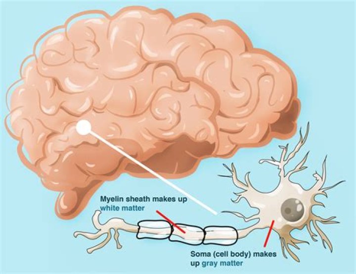
Myelin gives the white matter its color. It also protects the nerve fibers from injury. Also, it improves the speed and transmission of electrical nerve signals along extensions of the nerve cells called axons. By comparison, gray matter is tissue found on the surface of the brain (cortical)..
Simply so, what does it mean when you have white matter on the brain?
White matter disease is a disease that affects the nerves that link various parts of the brain to each other and to the spinal cord. These nerves are also called white matter. White matter disease causes these areas to decline in their functionality. This disease is also referred to as leukoaraiosis.
Furthermore, why is your brain darker on the outside than on the inside? So the cell body and the terminal appears dark gray, but the axon along its length appears light gray. So the human brain evolved with the cell bodies external and the connections internal. And so the grey matter is on the outside and the white matter is on the inside.
In respect to this, what is the difference between white matter and gray matter in the brain?
Grey matter is distinguished from white matter in that it contains numerous cell bodies and relatively few myelinated axons, while white matter contains relatively few cell bodies and is composed chiefly of long-range myelinated axons. The colour difference arises mainly from the whiteness of myelin.
Can stress cause white matter lesions?
Stress can damage the brain. Neuroscientists at a UC Berkeley lab have uncovered evidence that a well-known stress hormone trips a switch in stem cells in the brain, causing them to produce a white matter cell that ultimately can change the way circuits are connected in the brain.
Related Question Answers
Can you die from white matter disease?
White matter disease has been implicated in tissue and clinical outcomes of patients with acute ischemic stroke, and data link white matter disease burden measured semiquantitatively and functional dependence or death in patients with spontaneous primary brain hemorrhage, according to the investigators.What is white matter on MRI?
White matter hyperintensities (WMHs) are lesions in the brain that show up as areas of increased brightness when visualised by T2-weighted magnetic resonance imaging (MRI). WMH's are also referred to as Leukoaraiosis and are often found in CT or MRI's of older patients.At what age does white matter disease start?
Age-related changes in the brain -- the appearance, starting around age 60, of "white-matter lesions" among the brain's message-carrying axons -- significantly affect cognitive function in old age. White-matter lesions are small bright patches that show up on magnetic resonance imaging (MRI) of the brain.Is white matter disease the same as dementia?
White matter dementia (WMD) is a syndrome introduced in 1988 to highlight the potential of cerebral white matter disorders to produce cognitive loss of sufficient severity to qualify as dementia.Is white matter bad?
More evidence has been accumulated that damage to cognitive areas is widespread from white matter disease. White matter disease is responsible for about a fifth of all strokes worldwide, more than doubles the future risk of stroke, and is a contributing factor in up to 45% of dementias.Are white spots normal on brain MRI?
White Spots on a Brain MRI White spots may be described in your MRI report as high signal intensity areas (HSIA), white matter hyperintensities, leukoaraiosis (often used if spots are felt to be caused by decreased blood flow), or nonspecific white matter changes.Does white matter disease cause headaches?
Migraines Linked to Brain Lesions, White Matter Damage. Women are twice as likely as men to experience migraines, according to the U.S. Centers for Disease Control and Prevention . In the study, researchers used six population-based studies and 13 clinical studies to compare migraines' long-term effects.What parts of the brain are white matter?
White matter is found in the deeper tissues of the brain (subcortical). It contains nerve fibers (axons), which are extensions of nerve cells (neurons). Many of these nerve fibers are surrounded by a type of sheath or covering called myelin. Myelin gives the white matter its color.Why is white matter important?
The white matter is white because of the fatty substance (myelin) that surrounds the nerve fibers (axons). This myelin is found in almost all long nerve fibers, and acts as an electrical insulation. This is important because it allows the messages to pass quickly from place to place.Is Gray Matter good?
Grey matter is a diffuse network of brain regions thought to be involved in information processing. It is rich in nerve cell bodies and looks grey to the naked eye. They found that people with high IQ scores had significantly more grey matter in 24 of the regions than people with lower scores.How does white matter affect the brain?
This tissue contains millions of nerve fibers, or axons, that connect other parts of the brain and spinal cord and signal your nerves to talk to one another. A fatty material called myelin protects the fibers and gives white matter its color. When it becomes diseased, the myelin breaks down.How much of the brain is gray matter?
While 20 percent of all the oxygen we breathe goes directly to the brain, an overwhelming majority of that (95 percent) suffuses into the gray matter. Overall, gray matter occupies 40 percent of the cerebrum, while white matter fills the remaining 60 percent.Can white matter be repaired?
White matter injuries occur when white matter tracts (bundles of myelinated axons) are damaged. White matter injuries are very serious, but, depending on the type and extent of the injury, extensive recovery may occur. As long as the neuron cell bodies remain healthy, axons can regrow and slowly repair themselves.What does loss of gray white matter mean?
A system to assess and quantify the cerebral edema by studying the grey-to-white matter ratio (GWR) in the CT has been proposed (15). In these studies, the loss of the differentiation between grey matter and white matter on the brain CT was a marker of cerebral oedema in patients after cardiac arrest (18-21).What is the function of white matter related to memory?
White matter is made up of the neuronal axons that connect neurons in the "gray matter" brain regions. White matter also helps the regions of the brain to communicate with one another. "Historically, a lot of people have put their eggs in the gray matter basket.What is gray matter disease?
"Gray matter disease causes progressive symptoms, like fatigue and memory loss. These higher brain functions are called cognitive functions. Most MS disability actually comes from cognitive dysfunction."What color are brains?
gray
How are white and GREY matter arranged?
White matter is buried deep in the brain, while gray matter is mostly found on the brain's surface, or cortex. The spinal cord, which transmits nerve impulses to and from the rest of the body, has the opposite arrangement: gray matter at its core with insulating white matter on the outside.Where is gray matter located?
White matter is found buried in the inner layer of the brain's cortex, while the grey matter is mainly located on the surface of the brain. The spinal cord is arranged in the opposite way, with grey matter found deep inside its core and the insulating white matter wrapped around the outside.