There are two main types of light microscopes: COMPOUND and STEREO microscopes. COMPOUND MICROSCOPES are so called because they are designed with a compound lens system. The objective lens provides the primary magnification which is compounded (multiplied) by the ocular lens (eyepiece)..
Also question is, what is the difference between a compound microscope and a light microscope?
A compound light microscope is a microscope with more than one lens and its own light source. In this type of microscope, there are ocular lenses in the binocular eyepieces and objective lenses in a rotating nosepiece closer to the specimen.
Beside above, what is the purpose of a compound light microscope? Simple light microscopes use a single lens to magnify an object and cannot reach high magnification. Compound light microscopes use two sets of lenses - an objective lens and an eyepiece - to produce images.
Similarly, what does compound mean in microscope?
Compound microscope: A microscope that consists of two microscopes in series, the first serving as the ocular lens (close to the eye) and the second serving as the objective lens (close to the object to be viewed).
What is the correct path of light in a compound microscope?
(One light path, one lens = simple microscope) Compound refers to the fact that in order to enlarge an image, a single light path passes through a series of lenses in a line. Each lens magnifies the image over the previous one.
Related Question Answers
What are the types of compound microscope?
There are two main types of light microscopes: COMPOUND and STEREO microscopes. COMPOUND MICROSCOPES are so called because they are designed with a compound lens system. The objective lens provides the primary magnification which is compounded (multiplied) by the ocular lens (eyepiece).What are the parts of a compound light microscope?
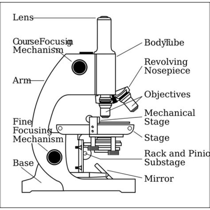
The three basic, structural components of a compound microscope are the head, base and arm. - Head/Body houses the optical parts in the upper part of the microscope.
- Base of the microscope supports the microscope and houses the illuminator.
- Arm connects to the base and supports the microscope head.
What are the parts and functions of a compound microscope?
Body tube (Head): The body tube connects the eyepiece to the objective lenses. Arm: The arm connects the body tube to the base of the microscope. Coarse adjustment: Brings the specimen into general focus. Fine adjustment: Fine tunes the focus and increases the detail of the specimen.What are the advantages of a compound microscope?
Thus, the total magnification in a compound microscope is a function of the objective magnification multiplied by the eyepiece magnification. The advantage of having a compound lens system as opposed to a simple magnifier is that much higher magnifications can be achieved with two lenses rather than one.What is the highest magnification of a light microscope?
Light microscopes combine the magnification of the eyepiece and an objective lens. Calculate the magnification by multiplying the eyepiece magnification (usually 10x) by the objective magnification (usually 4x, 10x or 40x). The maximum useful magnification of a light microscope is 1,500x.What type of microscope is used in schools?
High School Microscopes These are the most popular microscopes used in high schools around the world. High school compound microscopes will always have magnification of 40x, 100x, and 400x. Many of the high school microscopes will also have 1000x magnification.What is total magnification?
Total magnification is when the object being viewed is magnified to its maximum limit.Why must a specimen be centered?
The specimen must be centered in the field of view on low power before going to high power because on high power, it is difficult to find the specimen. Describe the changes in the field of view and the amount of available light when going from low to high power using the compound microscope. it becomes blurry.What is the principle of compound microscope?
A compound microscope works on the principle that when a tiny object to be magnified is placed just beyond the focus of its objective lens, a virtual, inverted and highly magnified image of the object is formed at the least distance of distinct vision from the eye held close to the eye piece.What can a compound microscope see?
With a limit of around 2000X magnification you can view bacteria, algae, protozoa and a variety of human/animal cells. Viruses, molecules and atoms are beyond the capabilities of today's compound microscopes and can be viewed only with an electron microscope.What is the magnification of a compound microscope?
Compound microscopes have a "nosepiece" with a rotating objective turret, which allows you to change the magnification level for different specimens. The standard objectives are 4x, 10x, and 40x for total magnification of 40x, 100x, and 400x. DIN is an international standard of lens quality.Who made the first compound microscope?
Hans Janssen
How is the image produced in a compound light microscope?
A compound microscope uses two or more lenses to produce a magnified image of an object, known as a specimen, placed on a slide (a piece of glass) at the base. The light rays hit an angled mirror and change direction, traveling straight up toward the specimen. The mirror pivots.What is the importance of microscope?
Microscopes are used in viewing the specimens that are relatively very small in size, they are used to view the cellular structures of organs, germs and bacteria, They play a very important role in laboratory for the tissues and organisms which are too small to be seen clearly with the naked eye.What is the resolution of a light microscope?
The maximum magnification of light microscopes is usually ×1500, and their maximum resolution is 200nm, due to the wavelength of light. An advantage of the light microscope is that it can be used to view a variety of samples, including whole living organisms or sections of larger plants and animals.What can you see at 1000x magnification?
At 1000x magnification you will be able to see 0.180mm, or 180 microns.What is the purpose of immersion oil in light microscopy?
In light microscopy, oil immersion is a technique used to increase the resolving power of a microscope. This is achieved by immersing both the objective lens and the specimen in a transparent oil of high refractive index, thereby increasing the numerical aperture of the objective lens.What is simple microscope?
A Simple microscope is a microscope that uses only one lens for magnification. It is the original design of the light microscope. Van Leeuwenhoek's microscopes consisted of a small single converging lens mounted on a brass plate with a screw mechanism to hold the sample or specimen to be inspected.What is the difference between 4x 10x and 40x on a microscope?
The same principle apply to stereo microscopes, a 10X eye piece combined with a 4X objective lens will produce 40X magnification. The total magnification will be 7.5X to 75X when combined with 10X ocular lens. The total magnification will be 18.75X to 187.5X when combined with a 25X ocular lens.