What kind of fracture is a complete craniofacial separation
By Daniel Johnston
A Le Fort III fracture is also called craniofacial separation because the craniofacial bone totally separates in a tripod pattern. This causes the nose and the dental arch to become mobile. The treatment of craniofacial separation (and other craniofacial injuries) is an individualised process.
What is a complete craniofacial separation called?
The fractures run parallel with the base of the skull, separating the entire midfacial skeleton from the cranial base. This discontinuity between the skull and the face is termed craniofacial dissociation.
What is a Lefort 4 fracture?
Le Fort fractures, also known as midaxillary fractures, are a group of fractures that affect the midface of the skull and collectively involve a partial or complete separation of the midface from the skull.
What is a Lefort 1 fracture?
Le Fort I level fractures are essentially a separation of the hard palate from the upper maxilla due to a transverse fracture running through the maxilla and pterygoid plates at a level just above the floor of the nose.What is a Le Fort 3 fracture?
A Le Fort III fracture includes fracture of the nasofrontal junction, bilateral fractures through the area of the frontozygomatic suture, and probable fractures of the zygomatic arch. These fractures are also referred to as craniofacial dysjunction.
What causes a Le Fort fracture?
Le Fort fractures account for 10-20% of all facial fractures. They result from exposure to a considerable amount of force. Motor vehicle accidents (MVAs) are the predominant cause; other causes include assaults and falls.
What type of fracture occurs through the medial and posterior lateral orbital walls?
Fractures of the orbital floor and the medial orbital wall (blowout fractures) are common midface injuries. Orbital fractures have a distinct trauma mechanism and are complex due to the complex anatomy of the bony and soft tissue structures involved.
Where is maxillary?
The maxilla is the bone that forms your upper jaw. The right and left halves of the maxilla are irregularly shaped bones that fuse together in the middle of the skull, below the nose, in an area known as the intermaxillary suture.How many types of Lefort fractures are there?
There are three types of Le Fort fractures.
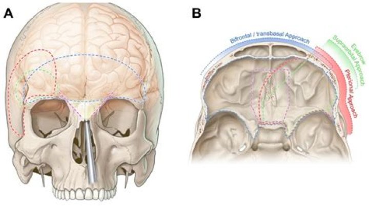
How do you assess Le Fort fracture?The level of a Le Fort fracture (ie, I, II, III) can often be determined by noting the structures of the midface that move in conjunction with the anterior maxilla. Illustration shows testing for mobility of the central midface. Illustration shows testing for mobility of the midface.
Article first time published onWhich bone is fractured in all types of Le Fort facial fractures?
Le Fort fractures are fractures of the midface, which collectively involve separation of all or a portion of the midface from the skull base. In order to be separated from the skull base, the pterygoid plates of the sphenoid bone need to be involved as these connect the midface to the sphenoid bone dorsally.
What is a Le Fort II fracture?
Definition. The Le Fort II fracture is also referred to as a pyramidal fracture. It commonly extends from the pterygoid plate through the maxilla, through the nasal orbital ethmoid area, and nasofrontal bone. Patients with Le Fort II injuries are often admitted to hospital unconscious and intubated.
What is an NOE fracture?
Naso-orbito-ethmoidal (NOE) fractures are complicated fractures of mid-face structure which include nasal, lacrimal, maxillary, frontal, and ethmoid bones. The central feature of NOE fracture is displacement of the medial orbital rim with the medial canthal ligament attached.
What is a LeFort procedure?
During the LeFort I surgery, the upper jaw (or maxilla) is separated from the rest of the face and repositioned. This repositioning of the bones of the face is also called orthognathic surgery. Once separated, the upper jaw can be moved up, down, forward, backward, tilted, or turned.
What is a Zygomaticomaxillary complex fracture?
A zygomatic complex fracture is a fracture that involves the zygoma and its surrounding bones. The typical lines of a zygomatic complex fracture are: A fracture emanating from the inferior orbital fissure superiorly along the sphenozygomatic suture to the frontozygomatic suture where it crosses the lateral orbital rim.
What does LeFort mean?
French and English: from Old French fort ‘strong’, ‘brave‘ (see Fort), with the definite article le.
What is medial orbital wall fracture?
Medial orbital wall blow out fractures, by definition is a pure internal fracture confined to the orbital wall without involvement of orbital rim. Two theories have been proposed to explain how these fractures occur, the hydraulic or buckling mechanisms.
What is orbital wall fracture?
Orbital fractures are breaks in any of the bones surrounding the eye area (also known as the orbit or eye socket). These fractures are almost always a result of a blunt force trauma injury, whether by accident or from sports.
What are orbital fractures?
An orbital fracture occurs when one or more of the bones around the eyeball break, often caused by a hard blow to the face. To diagnose a fracture, ophthalmologists examine the eye and surrounding area. X-ray and computed tomography scans may also be taken.
What types of facial fractures are likely to show the presence of leaking CSF?
Clinical Pathophysiology of Traumatic CSF Leak The most common fracture sites leading to CSF leaks following TBI are the frontal sinus (30.8%), sphenoid sinus (11.4–30.8%), ethmoid (15.4–19.1%), cribriform plate (7.7%), frontoethmoid (7.7%) and sphenoethmoid (7.7%).
Which complication is associated with a perforated globe?
There are some postoperative complications in ruptured globe injuries, such as a secondary cataract, corneal belted degeneration, iris atrophy, and an irregular pupil. Long-term contact between the silicone oil and lens is another factor for a secondary cataract.
How do you treat a maxillary fracture?
For all maxillary fractures, suspension of the soft tissue of the midface should be performed prior to closing the intraoral incisions with 3-0 chromic suture and closing the skin incisions with absorbable subcutaneous sutures and permanent skin sutures. Bicoronal flaps may be closed with skin staples.
Which type of fracture is most likely to cause trismus?
A posterior mandibular buttress fracture, especially when associated with a displaced fracture of the condylar process or dislocation of the temporomandibular joint, can cause malocclusion and trismus.
What kind of bone is maxilla?
The answer to the question, “What type of bone is the maxilla bone?” is simple – it is an irregular facial bone. You can refer to the maxilla bone as a single unit or as two paired but fused bones.
Is maxilla same as maxillary bone?
The two maxilla or maxillary bones (maxillae, plural) form the upper jaw (L., mala, jaw). Each maxilla has four processes (frontal, zygomatic, alveolar, and palatine) and helps form the orbit, roof of the mouth, and the lateral walls of the nasal cavity.
Which part of the maxillary bones form the roof of the mouth?
Terms in this set (5) Which part of the maxillary bones form the roof of the mouth? The palatine processes of the maxillary bones fuse on midline at the intermaxillary suture, forming the anterior portion of the hard palate (roof of the mouth).
What is blowout fracture?
A blowout fracture is a break of one or more of the bones that surround the eye. When an object strikes the eye, the force is transmitted into the eye compartment (orbit) [see figure 1], and the thinnest bones within the orbit will buckle or break from the force of the trauma.
What type of facial fracture is associated with the leakage of cerebrospinal fluid into the nasal sinus?
Frontal sinus fractures that extend through the posterior sinus wall can create a communication with the anterior cranial fossa resulting in leakage of cerebrospinal fluid, intracranial bleeding.
What is tripod fracture?
tri·pod frac·ture. a facial fracture involving the three supports of the malar prominence, the arch of the zygomatic bone, the zygomatic process of the frontal bone, and the zygomatic process of the maxillary bone.
How do you fix a broken Noe?
NOE fractures often result in a lateral splaying of the medial canthi, resulting in telecanthus. This can often be corrected by placement of a transnasal wire. When confronted with a NOE fracture requiring a transnasal wire, it is important to place the wire fixation in its proper posterior position.
What is a fractured Zygoma?
From Wikipedia, the free encyclopedia. A zygoma fracture (zygomatic fracture) is a form of facial fracture caused by a fracture of the zygomatic bone. A zygoma fracture is often the result of facial trauma such as violence, falls or automobile accidents.