What does hematoxylin and eosin stain
By Matthew Harrington
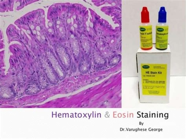
The H&E stain provides a comprehensive picture of the microanatomy of organs and tissues. Hematoxylin precisely stains nuclear components, including heterochromatin and nucleoli, while eosin stains cytoplasmic components including collagen and elastic fibers, muscle fibers and red blood cells.
What Colour does hematoxylin and eosin stain?
Hematoxylin has a deep blue-purple color and stains nucleic acids by a complex, incompletely understood reaction. Eosin is pink and stains proteins nonspecifically. In a typical tissue, nuclei are stained blue, whereas the cytoplasm and extracellular matrix have varying degrees of pink staining.
What is haematoxylin used to stain?
Principally used as a nuclear stain (to stain the cell nucleus), haematoxylin will also stain rough endoplasmic reticulum, ribosomes, collagen, myelin, elastic fibers, and acid mucins.
What is eosin staining used for?
Eosin can be used to stain cytoplasm, red blood cells, collagen, and muscle fibers for histological examination. It is most often used as a counterstain to hematoxylin in H&E staining.What cell structures did eosin stain?
Eosin stains the cytoplasm besides the RER. Cells with numerous mitochondria (e.g., ductal cells, osteoclasts, muscle cells and parietal cells) stain particularly intensely with eosin.
Why is hematoxylin and eosin staining used routinely in histopathology?
Hematoxylin and Eosin (H&E) staining is used routinely in histopathology laboratories as it provides the pathologist/researcher a very detailed view of the tissue. It achieves this by clearly staining cell structures including the cytoplasm, nucleus, and organelles and extra-cellular components.
What does trichrome stain?
Trichrome staining is used to visualize connective tissues, particularly collagen, in tissue sections. In a standard Masson’s Trichrome procedure, collagen is stained blue, nuclei are stained dark brown, muscle tissue is stained red, and cytoplasm is stained pink.
How is hematoxylin made?
Hematoxylin is a natural product extracted from the heartwood of the logwood tree (Haematoxylum campechianum).What is the difference between hematoxylin and eosin?
Hematoxylin and eosin are important dye compounds in staining microstructures such as proteins in the cytoplasm. The key difference between hematoxylin and eosin is that hematoxylin is a basic dye, whereas eosin is an acidic dye.
Is hematoxylin a dye?Hematoxylin is a naturally occurring chemical used as the basis of a dye in laboratories throughout the world to stain nuclei in microscope slide preparations. … Hematoxylin remains the most popular nuclear stain in histology.
Article first time published onWhat does hematoxylin and eosin not stain?
Results. Hematoxylin principally colors the nuclei of cells blue or dark-purple, along with a few other tissues, such as keratohyalin granules and calcified material. Eosin stains the cytoplasm and some other structures including extracellular matrix such as collagen in up to five shades of pink.
How does mitochondria stain with hematoxylin and eosin?
On histological slides stained with hematoxylin and eosin (H&E), the color of cytoplasm in cells containing many mitochondria is: red or pink. blue or purple.
Is hematoxylin acidic or basic?
Haematoxylin can be considered as a basic dye. It is used to stain acidic structures a purplish blue. DNA in the nucleus, and RNA in ribosomes and in the rough endoplasmic reticulum are both acidic, and so haemotoxylin binds to them and stains them purple.
What is stain principle of hematoxylin and eosin procedure clinical application of stain?
The principle behind H & E stain is the chemical attraction between tissue and dye. Hematoxylin, a basic dye imparts blue-purple contrast on basophilic structures, primarily those containing nucleic acid moeties such as chromtatin, ribosomes and cytoplasmic regions rich in RNA.
Is hematoxylin a fluorescent?
Hematoxylin has broad absorption between 400 and 700 nm, with virtually no fluorescence emission. …
How do you make a hematoxylin stain?
- Boil 800 mL water and add Potash alum till it is dissolved.
- Mix 4 grams hematoxylin in 60 mL ethanol. Shake well to dissolve it.
- When potash is dissolved now add the solution of hematoxylin + ethanol solution.
What is the meaning of trichrome?
Medical Definition of trichrome : coloring tissue elements differentially in three colors a trichrome biological stain.
What does Ponceau Fuchsin stain?
The ponceau-fuchsin counterstain gives good differentiation be- tween muscle fibers (which are stained bright red) and collagen con- nective tissue fibers (dull pink); it stains more crisply and brightly than eosin and is equally simple to use.
What stains blue in trichrome?
Masson’s trichrome. Nuclei and other basophilic (basic-liking) structures are stained blue, cytoplasm, muscle, erythrocytes and keratin are stained bright-red. Collagen is stained green or blue, depending on which variant of the technique is used.
Why are histological stains important?
Staining is used to highlight important features of the tissue as well as to enhance the tissue contrast. Hematoxylin is a basic dye that is commonly used in this process and stains the nuclei giving it a bluish color while eosin (another stain dye used in histology) stains the cell’s nucleus giving it a pinkish stain.
What are the three major groups of stains used for histopathology?
- Routine stains. Haematoxylin & Eosin.
- Special stains. Van Gieson. Toluidine Blue. Alcian Blue. Giemsa. Reticulin. Nissl. Orcein. Sudan Black B. Masson’s Trichrome. Mallory’s Trichrome. Azan Trichrome. Cason’s Trichrome. Periodic Acid Schiff. Weigert’s Resorcin Fuchsin.
Why are special stains used?
“Special stains” are processes that generally employ a dye or chemical that has an affinity for the particular tissue component that is to be demonstrated. They allow the presence/or absence of certain cell types, structures and/or microorganisms to be viewed microscopically.
How do you prepare hematoxylin and eosin stain?
Method – Dissolve the hematoxylin in absolute alcohol and ammonium alum in hot water. Mix the two solutions and heat to boiling. Remove from flame, and add mercuric oxide and cool rapidly. Glacial acetic acid if added gives brisk nuclear staining, but life of the solution is reduced.
What are histological features?
Histology, also known as microscopic anatomy or microanatomy, is the branch of biology which studies the microscopic anatomy of biological tissues. Histology is the microscopic counterpart to gross anatomy, which looks at larger structures visible without a microscope.
Is H&E an IHC?
H&E also serves as what is arguably the most popular background stain in immunohistochemistry (IHC). When using an antibody to detect a specific protein through IHC, a background stain such as H&E is used to simultaneously visualize the cells where the protein is being detected.
Where is hematoxylin found?
Hematoxylin is a basic dye derived from the heartwood of Palo de Campeche ( Haematoxylum campechianum), the logwood tree native to Mexico and Central America.
What is the pH of hematoxylin?
The pH and peak of absorbance of the aliquots were pH = 2.0 450 NM, 2.5 505, 2.6 507, 2.7 515, 2.8 520, 2.9 530, 3.0 540, 3.1 550, 3.3 560, 3.5 560. In the stained material in the intensity of nuclear staining was about the same at all pH levels but non-specific staining was greatest in slides stained at pH = .
What is hematoxylin in histopathology?
Hematoxylin and eosin (H&E) is the most widely used stain in histology and allows localization of nuclei and extracellular proteins. Hematoxylin, not a dye itself, produces the blue Hematin via an oxidation reaction with nuclear histones causing nuclei to show blue.
Is hematoxylin positive or negative?
Haematoxylin in complex with aluminium salts is cationic and acts as a basic dye. It is positively charged and can react with negatively charged, basophilic cell components, such as nucleic acids in the nucleus. These stain blue as a result.
Is hematoxylin toxic?
Ingestion Toxic if swallowed. May be harmful if absorbed through skin. Causes skin irritation. Eyes Causes eye irritation.
Why is hematoxylin basic?
(Haematoxylin is not strictly a basic dye, but it is used with a ‘mordant’ that makes this stain act as a basic dye. The mordant (aluminium salts) binds to the tissue, and then haematoxylin binds to the mordant, forming a tissue-mordant-haematoxylin linkage.)