A dull tympanic membrane (dull ear drum) occurs when something is wrong with the middle ear, such as an infection, inconsistent pressure in the ear,.
In respect to this, what does a healthy tympanic membrane look like?
1) Color/shape-pearly grey, shiny, translucent, with no bulging or retraction. 2) Consistency - smooth.
Furthermore, what is a normal tympanic membrane? The normal tympanic membrane is a pale, gray, ovoid, semi-transparent membrane located at the end of the external auditory canal.
Regarding this, what color is a normal tympanic membrane?
A normal TM is a translucent pale gray. An opaque yellow or blue TM is consistent with MEE.
What does a cloudy eardrum mean?
An ear infection is an infection of the middle ear (the space behind the eardrum). It is most often caused by bacteria. In 5% to 10% of children, the pressure in the middle ear causes the eardrum to rupture and drain a yellow or cloudy fluid. This small hole usually heals over the next few days.
Related Question Answers
What is the most common cause of a ruptured tympanic membrane?
ear infection
How do you fix a tympanic membrane?
Patch the eardrum with a piece of the patient's own tissue taken from a vein or muscle sheath (called tympanoplasty). This procedure will usually take 2 to 3 hours. Remove, replace, or repair 1 or more of the 3 little bones in the middle ear (called ossiculoplasty).What does the tympanic membrane do?
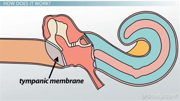
Tympanic membrane, also called eardrum, thin layer of tissue in the human ear that receives sound vibrations from the outer air and transmits them to the auditory ossicles, which are tiny bones in the tympanic (middle-ear) cavity.What does cone of light mean?
The cone of light, or light reflex, is a visible phenomenon which occurs upon examination of the tympanic membrane with an otoscope. Shining light on the tympanic membrane causes a cone-shaped reflection of light to appear in the anterior inferior quadrant.What color is an infected eardrum?
Ear Infection Diagnosis A normal, healthy eardrum has a pinkish-gray color as shown here. The healthy eardrum is clear, while an infected eardrum is bulging (swollen) and reddened. A doctor may also perform a tympanometry, which measures how the eardrum responds to a change of air pressure inside the ear.Can scar tissue on Eardrum be removed?
Scar tissue on the ear drum is usually caused by repeated ear infections. The scar tissue can lead to difficulty hearing and eventually ruptured ear drums. Surgery is one means of avoiding progressive hearing loss or ear drum rupture. There are no evaluations for Scar tissue removal (eardrum).Can you see the tympanic membrane?
Tympanic membrane perforations can usually be diagnosed by routine examination of the ear with an otoscope. Occasionally, wax or drainage may occlude the ear canal so that the eardrum can not be seen. Larger perforations or those that are around the ossicles cause larger degrees of hearing loss.What does a doctor see when he looks in your ear?
An otoscope or auriscope is a medical device which is used to look into the ears. Health care providers use otoscopes to screen for illness during regular check-ups and also to investigate ear symptoms. An otoscope potentially gives a view of the ear canal and tympanic membrane or eardrum.What does an ear infection look like?
Signs of Infection Here are some things to look for: A red, bulging eardrum. Clear, yellow, or greenish fluid behind the eardrum. There may also be some blood.Why is the tympanic membrane important?
The tympanic membrane's function is to assist in human hearing. When sound waves enter the ear, they strike the tympanic membrane. The membrane vibrates with the force of the sound wave strike and transmits the vibrations further in, to the bones of the middle ear.What does a bulging tympanic membrane indicate?
The classic findings of acute otitis media, such as fever and earache, are sometimes absent even in cases confirmed by tympanocentesis. A bulging, red, immobile tympanic membrane is highly associated with acute otitis media. However, many physicians rely on redness of the eardrum as the main diagnostic clue.What does a white tympanic membrane mean?
Myringosclerosis and tympanosclerosis are similar conditions which affect the middle ear, causing the eardrum to appear bright white. The whiteness is due to calcium deposits which form on the tympanic membrane, which is more commonly called the eardrum. Tympanosclerosis can cause symptoms such as hearing loss.Is presbycusis normal?
Presbycusis is the loss of hearing that gradually occurs in most individuals as they grow older. Hearing loss is a common disorder associated with aging. About 30-35 percent of adults age 65 and older have a hearing loss. It is estimated that 40-50 percent of people 75 and older have a hearing loss.How do you drain fluid from your middle ear?
One form of direct treatment is ear tubes, which help drain fluid from behind the ears. Removing the adenoids can also help treat or prevent OME in some children. When adenoids become enlarged they can block ear drainage.Can a ruptured tympanic membrane heal?
A ruptured eardrum can result in hearing loss. It can also make your middle ear vulnerable to infections. A ruptured eardrum usually heals within a few weeks without treatment. But sometimes it requires a patch or surgical repair to heal.Where is the cone of light located in the ear?
You can see the tiny bones of the middle ear pushing on the eardrum. You see a cone of light, known as the "light reflex," reflecting off the surface of the eardrum. This cone of light is at the 5 o'clock position in the right ear and at the 7 o'clock position in the left ear.What is serous otitis media in adults?
Serous otitis media is fluid trapped behind your tympanic membrane (eardrum), without an ear infection. Your eardrum is in your middle ear. Serous otitis media is also called otitis media with effusion. You may have fluid in your ear for months, but it usually goes away on its own. The fluid may be in one or both ears.How do you examine an ear drum?
For an ear exam, the doctor uses a special tool called an otoscope to look into the ear canal and see the eardrum. Your doctor will gently pull the ear back and slightly up to straighten the ear canal. For a baby under 12 months, the ear will be pulled downward and out to straighten the ear canal.Why is my eardrum blue?
The "blue ear drum" generally refers to a condition in which blood or blood products are found in the middle ear. After all possible causes for hemotympanum, including blood dyscrasias and trauma are searched for and ruled out, the patient may have chronic serous otitis media accompanied by bloody effusion.