Smears in the gels could be a result of aggregates produced during the heating process. Although SDS-PAGE method unfolds and protects the protein from aggregation, the heating process sometimes produces "unbreakable" aggregates" with variable sizes that enter the gel and create a smear..
Similarly, you may ask, what causes smearing in gel electrophoresis?
Gel electrophoresis allows scientists to visualize digested samples and measure the sizes of the fragments. Smearing results from improperly prepared agarose gels, loading an undiluted sample into the wells or using poor quality samples.
Also, why is SDS added to the sample and the gel? SDS-PAGE separates proteins primarily by mass because the ionic detergent SDS denatures and binds to proteins to make them uniformly negatively charged. Thus, when a current is applied, all SDS-bound proteins in a sample will migrate through the gel toward the positively charged electrode.
People also ask, how can I improve my SDS PAGE results?
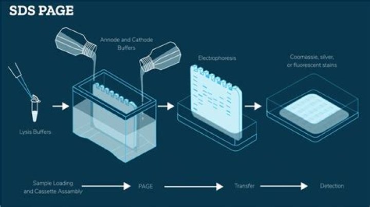
Five Tips for Good Bands with SDS PAGE Protein Gels
- Consider Heating and / or Using Reducing Agents to Prepare Samples.
- Rinse and Fill Wells with Water.
- Load any Empty Wells with Sample Buffer.
- Run Straight Away After Loading Samples.
- Keep Gel Temperature Low and Even.
Why SDS PAGE is vertical?
The first reason is that SDS-PAGE gels have two component gels – the stacking gel and the resolving gel. The vertical system allows you to make them sequentially. The second reason is that oxygen inhibits the polymerization of SDS-PAGE gels.
Related Question Answers
What is a DNA smear?
A smear along the path of nucleic acid movement is simply many bands that cannot be easily distinguished. For example, sizes of molecules in genomic DNA or degraded RNA vary extensively so they are manifested as smears.How do you prevent smearing in gel electrophoresis?
Gel preparation - Clean the gel comb properly before using it in casting the gel.
- To prevent sample leakage through the bottom of the gel and smearing of the sample bands, do not push the comb all the way to the bottom of the horizontal gel.
- Avoid overfilling the gel tray, as this can result in connected wells.
How can you tell if your gel is running properly?
How can one tell if their gel electrophoresis is running properly? It bubbles. You can see the methyl blue move from the well into the gel. The DNA runs to red.Why do PCR products smear?
Why do I get smeared PCR products? Set up all reaction mixtures in an area separate from that used for DNA preparation or PCR product analysis. enzyme concentration too high. When using HotStarTaq or Taq DNA Polymerase, use 2.5 units per 100 µl reaction.Why does gel electrophoresis not work?
You may have ran your DNA out of the gel by running for too long. You may have melted your gel by running for too long or using a stale electrophoresis buffer with a low buffer capacity. You may have used a terribly wrong percentage of agarose and DNA either stuck in the well or prematurely ran out of the gel.Why are there two bands in gel electrophoresis?
The gel matrix acts as a sieve: smaller DNA molecules migrate faster than larger ones, so DNA molecules of different sizes separate into distinct bands during electrophoresis. So, for example, 50ng of DNA in a band gives two times more (= brighter) staining than 25ng.Why is ethidium bromide used in gel electrophoresis?
Ethidium Bromide (EtBr) is sometimes added to running buffer during the separation of DNA fragments by agarose gel electrophoresis. It is used because upon binding of the molecule to the DNA and illumination with a UV light source, the DNA banding pattern can be visualized.How do you troubleshoot PCR?
Use the lowest possible concentration when appropriate. Adjust the annealing temperatures, as high concentrations of PCR additives or co-solvents weaken primer binding to the target. Increase the amount of DNA polymerase, or use DNA polymerases with high processivity.How do I run SDS gel?
Turn on the power supply. Run the gel at a constant voltage of 120-150 V. Run the gel until the blue dye front nearly reaches the bottom of the gel. This may take between 45-60 min.How much protein should I load on an SDS gel?
A rule of thumb for mini-slab gels is to load about 0.5 microgram protein per expected band.How long should I run SDS PAGE?
Typical conditions include runs at 100-150 volts for 40-60 minutes or until the dye front has reached the bottom of the gel. Letting it run too long will result in losing your lower molecular weight bands.Why is it important to denature proteins before electrophoresis?
Denaturing the proteins nullifies structural effects on mobility, allowing separation on a true charge/mass ratio basis. It also separates subunits in multimeric proteins, allowing analysis of large, complex aggregates. SDS is the most commonly used detergent in protein electrophoresis.How much protein should I load for a well?
Cell lysates, membrane and nuclear lysates: load 20 to 30 µg of total protein per well. This may require some optimization depending on the expression level of the protein in the sample you are testing. Purified protein (recombinant or endogenous): load 10 to 100 ng of protein.How do you make a protein sample for SDS PAGE?
For a 2x sample buffer use equal amounts of sample and buffer, for 5x sample buffer use 4 parts of sample and 1 part of buffer (for examle 40µl + 10µl). Heat the mixed samples for 5 minutes at 95°C, cool them immediately on ice and load an appropriate amount of it on the gel.How much protein should I load for Coomassie?
For standard Coomassie staining, I generally load about 2ug of a singular purified protein per well. You should get a crisp dark band. In my experience, if you go much over 5ug your band will start to become more blob and less band.Why are the protein samples heated before loading on the gel?
Heat ensures that your samples are truly denatured. In addition, heat loosens up samples gummy from DNA and cellular debris, making the samples easier to load. So that's the long answer as to why you heat protein samples prior to loading.What is the principle of SDS PAGE?
SDS-PAGE is an electrophoresis method that allows protein separation by mass. The medium (also referred to as ′matrix′) is a polyacrylamide-based discontinuous gel. In addition, SDS (sodium dodecyl sulfate) is used. About 1.4 grams of SDS bind to a gram of protein, corresponding to one SDS molecule per two amino acids.What is SDS function?
Sodium Dodecyl Sulfate, Molecular Biology Grade (SDS), is a detergent that is known to denature proteins. It is used in denaturing polyacrylamide gel electrophoresis for the determination of protein molecular weight.Why is pH important in SDS PAGE?
The main reason is to differentiate the rate of migration while the proteins are stacking into a tight band in the wells, before they enter resolving gel for separation. The respective pH influences the charge of ions in the running buffer, and thus their migration when electric current is turned on.