What are the five components of intraoral film
By Sophia Dalton
film base.thin adhesive layer.gelatin.silver halide crystals.protective layer.
Which is a component of intraoral film?
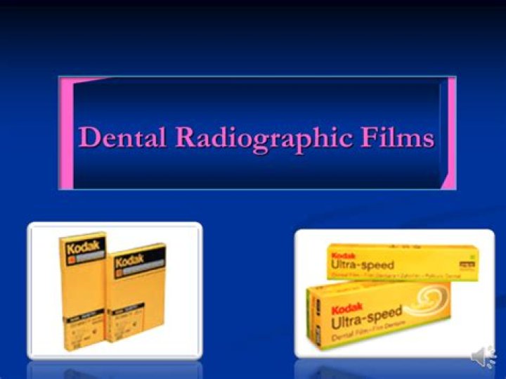
Intraoral dental film is made up of a semiflexible, clear cellulose acetate film base that is coated on both sides with an emulsion of silver bromide, silver halide, and silver iodide that are sensitive to radiation.
What are the parts of a dental film?
- i. Film base. • The film base is a flexible piece of polyester plastic (polyethylene terephthalate) 0.2 mm in thickness. …
- ii. Adhesive layer. • …
- iii. Film emulsion. • …
- iv. Protective layer. •
What are the five basic sizes of intraoral dental film?
- Child (size 0)
- Narrow anterior (size 1)
- Adult size (size 2)
- Preformed bite-wing (size 3)
- Occlusal (size 4)
What are the two types of extraoral film?
- Panoramic X-rays show the entire mouth area — all the teeth in both the upper and lower jaws — on a single X-ray. …
- Tomograms show a particular layer or “slice” of the mouth and blur out other layers. …
- Cephalometric projections show an entire side of the head.
How many components are there in a dental film composition?
Composition. X-ray film has two principal components: (1) emulsion and (2) base. The emulsion, which is sensitive to x rays and visible light, records the radiographic image. The base is a plastic supporting material onto which the emulsion is coated (Fig.
What is intraoral film used for?
Types of Intraoral X-Rays Bite-wing X-rays are used to detect decay between teeth and changes in bone density caused by gum disease. They are also useful in determining the proper fit of a crown (or cast restoration) and the marginal integrity of fillings.
Which of the following components of the film packet reduces scatter radiation?
The purpose of the lead foil in the film packet is to absorb back-scattered x-rays to reduce film fog. Dental film emulsion is about 90 to 99 percent silver bromide and 1 to 10 percent silver iodide.How do extraoral and intraoral film differ?
There are two main types of dental X-rays: intraoral (meaning the X-ray film is inside the mouth) and extraoral (meaning the X-ray film is outside the mouth). Intraoral X-rays are the most common type of dental X-ray taken. … Extraoral X-rays show teeth, but their main focus is the jaw and skull.
How many items compose an intraoral film packet?List and describe the four parts of an intraoral film packet. The four parts of the intraoral film are film, black paper wrapping, lead foil, and moisture resistant out wrapping.
Article first time published onWhat are the components of Xray film?
X-ray films for general radiography consist of an emulsion-gelatin containing radiation sensitive silver halide crystals, such as silver bromide or silver chloride, and a flexible, transparent, blue-tinted base.
What is the largest intraoral film size?
This bitewing film shows all of the posterior teeth on one side of the arch in one radiograph. The occlusal film is the largest intraoral film and is almost four times as large as a standard Size 2 periapical film (57×76 mm). Size 4: This occlusal film is used to show large areas of the upper or lower jaw (Fig. 11.4).
What is the most common film size for an intraoral image?
Place a bite tab on the white side of a Size #2 intraoral film. Intraoral films are available in different sizes; the #2 is the most commonly used size for BWs. Common errors: Placing the bite tab on the incorrect side of the film places it into the mouth backwards, producing an image that is reversed.
What are the component parts of an automatic film processor?
Automatic film processors, sometimes called film processing systems, typically have six main subsystems: film transport, temperature, circulation, replenishment, drying and electrical control.
What does intraoral mean?
Intraoral: Within the mouth.
What does intraoral periapical mean?
The American Dental Association defines a bitewing radiograph as “interpoximal view radiograph of the coronal portion of the tooth” and a periapical radiograph as “a radiograph made by the intraoral placement of film for disclosing the apices of the teeth.”
What are the three types of intraoral radiographs?
There are three types of diagnostic radiographs taken in today’s dental offices — periapical (also known as intraoral or wall-mounted), panoramic, and cephalometric.
What are the different types of film in radiography?
The film typically used for the intraoral bitewing exam falls into three film speed classes – D (slowest), E and F-speed (fastest). Like photographic film, the faster the film, the less exposure it needs. Film speed can be an important aspect in determining the amount of radiation exposure received by a patient.
What is Movie composition?
Composition refers to how the elements on screen (actors, scenery, props, etc.) appear in respect to each other and within the frame itself. In the earliest days of cinema, film composition basically mimicked that of a stage play. Directors staged all actors and important information to face the audience.
What type of film is used for duplication?
Duplicating film is single emulsion film. In the darkroom, the one side of the film will appear lighter than the other side of the film. The lighter side of the film must be placed toward the light in the duplicating machine. Light to light.
Which type of film is more sensitive to light radiation intraoral or extraoral?
A screen film is more sensitive to fluorescent light than to direct exposure to X-rays. Nonscreen extraoral film is commonly used in extraoral radiography.
What type of photographic film is used to make an identical copy of an intraoral or extraoral radiograph?
The duplicating Film is a type of film that is used to make an identical copy of an intraoral or extraoral radiograph.
Which type of intraoral projection is best for visualizing interproximal surfaces for decay?
In carious prone patients radiograph should be taken every 6 – 12 months; in non-caries prone patients every 18 – 24 months is adequate. The best radiographic view for visualizing both interproximal caries and periodontal bone height is bite-wing radiographs.
What is a film packet?
a thin sheet of material (e.g., gelatin, cellulose acetate) specially treated for use in photography or radiography; used also to designate the sheet after exposure to the energy to which it is sensitive.
How does a film holder protect the patient from unnecessary radiation?
How do positioning instruments protect the patient from unnecessary radiation? It keeps the patients hands and fingers from being exposed to x-radiation. Bite block with a backing plate and a slot for film sensor retention.
What hardens the film emulsion during film processing?
The fixer hardens the film emulsion during the process. When a beam of photons exposes an x-ray film, it chemically changes the photosensitive silver halide crystals in the film emulsion (latent image). Important: Exposed areas will become radiolucent, whereas unexposed areas will become radiopaque.
What is the main substance contained in the film emulsion covering the film?
The film’s emulsion layer contains silver halide crystals that capture the x-ray image. Small crystal grains of silver halide (1.0 to 1.5 microns in diameter) comprise the light sensitive substance in the emulsion.
How latent image is formed?
A latent image is an invisible image produced by the exposure to light of a photosensitive material such as photographic film. When photographic film is developed, the area that was exposed darkens and forms a visible image. … If intense exposure continues, such photolytic silver clusters grow to visible sizes.
What is the difference between radiopaque and radiolucent?
Radiolucent – Refers to structures that are less dense and permit the x-ray beam to pass through them. Radiolucent structures appear dark or black in the radiographic image. Radiopaque – Refers to structures that are dense and resist the passage of x-rays.
What determines film speed?
The film speed is determined by: the size of its silver halide crystals; the thickness of the emulsion; and the presence of radiosensitive dyes. Films are described as “fast” if the film requires little radiation to produce an image and “slow” if more radiation is needed.
Which type of exposure is a panoramic?
Panoramic dental x-ray uses a very small dose of ionizing radiation to capture the entire mouth in one image. It is commonly performed by dentists and oral surgeons in everyday practice and may be used to plan treatment for dentures, braces, extractions and implants.